Tamura,
R., Eifuku, S., Uwano, T., Sugimori, M., Uchiyama, K., Ono, T. A method for recording
evoked local field potentials in the primate dentate gyrus
in vivo. Hippocampus, 21(5): 565-574, 2011 [査読有]
Recording evoked local field potentials
(LFPs) in the hippocampus in vivo has yielded
us useful information about the neural mechanisms of learning and memory.
Although this technique has been used in studies of the hippocampus of rodents,
lagomorphs and felines, it has not yet been applied to the primate hippocampus.
Here we report a method for recording evoked LFPs in
the hippocampus of monkeys. A stimulation electrode and a recording electrode
were implanted in the perforant pathway and dentate gyrus, respectively, under the guidance of
electrophysiological recording. With a low stimulus intensity just above the
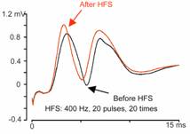
threshold, the potential appeared as a slow positive-wave component, which was
regarded as field excitatory postsynaptic potential (putative fEPSP); as stimulation intensity increased, the fEPSP amplitude increased, followed by a sharp negative
component which was regarded as putative population spike. When the
coordinates of the recording or stimulation electrode were moved stepwise, we
observed a systematic change in the waveforms of evoked LFPs;
this change corresponded to the structural arrangement through which the
electrode passed. In a test for short-term synaptic plasticity by paired-pulse
stimulation, potentials evoked by the second pulse were influenced by the first
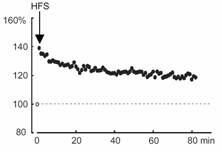
one in a manner dependent on inter-pulse intervals. In a test for long-term
synaptic plasticity by high-frequency stimulation, the slopes of the fEPSPs and the area of population spikes were increased for
more than one hour. These results indicate that the method developed in the
present study is useful for testing theories of hippocampal
functions in primates.
KEY
WORDS: perforant pathway; field excitatory
postsynaptic potential; population spike; synaptic plasticity; monkeys
(抄録和訳)
インビボで海馬から誘発局所電場電位を記録することにより、学主記憶の神経機構に関する有用な情報を得ることができる。この手法は、げっ歯類、ウサギやネコに適用されてきたが、霊長類動物の海馬にはいまだに用いられていない。本研究でわれわれは、サルの海馬で誘発電位を記録する方法について報告する。電気生理学的記録のガイド下に、刺激電極と記録電極を、それぞれ、貫通路と歯状回に埋め込んだ。閾値よりわずかに高い強度の刺激で、緩徐な要請は成分が出現したが、これはフィールド興奮性後シナプス電位(fEPSP)と思われる。刺激強度を増加するにつれて、fEPSPの振幅は増加し、それに引き続いて集合スパイク電位と思われる鋭い陰性波成分が出現した。段階的に刺激電極や記録電極の座標を移動すると、誘発電場電位波形の系統的な変化が観察された。この変化は、これら電極が通過する構造的(解剖的)な配置に一致していた。対パルス刺激により短期シナプス可塑性を検討してみたところ、第二刺激により誘発される電位は、パルス間隔に依存したかたちで第一刺激により影響された。高頻度刺激により長期シナプス可塑性を検討してみたところ、fEPSPのスロープと集合スパイク波形面積が一時間以上持続して増大していた。これらの結果は、本研究で開発した方法が、霊長類での海馬機能に関する仮説を検証するのに有用であることを示している。
キーワード:貫通路、フィールド興奮性シナプス後電、集合スパイク、シナプス可塑性、サル
本研究は、霊長類でもインビボで、海馬に長期増強を誘導できることを実証した世界で初めての研究である。